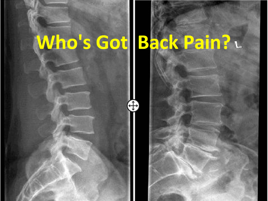

Diagnostic imaging in patients with back pain and/or leg pain is often used to assess nerve root compression due to disc herniation or spinal stenosis and cauda equina syndrome. However, the diagnostic accuracy of both history taking and physical examination is still insufficient. Clinical guidelines recommend history taking and physical examination to rule out LDH diagnosis. LDH is the most common spine disorder requiring surgical intervention. The quality of evidence was moderate to very low.Īpproximately 5–15% of patients with low back pain suffer from lumbar disc herniation (LDH). The summary estimates for MRI and myelography were comparable with CT (sensitivity: 81.3% (95%CI 72.3–87.7%) and specificity: 77.1% (95%CI 61.9–87.5%)). The prior probability of LDH varied from 48.6 to 98.7%. Nine studies investigated Computed Tomography (CT), eight myelography and six Magnetic Resonance Imaging (MRI).

We found 14 studies, all but one done before 1995, including 940 patients. We calculated summary estimates of sensitivity and specificity using bivariate analysis, generated linked ROC plots in case of direct comparison of diagnostic imaging tests and assessed the quality of evidence using the GRADE-approach. Two review authors independently selected studies, extracted data and assessed risk of bias. We aim to summarize the available evidence on the diagnostic accuracy of imaging (index test) compared to surgery (reference test) for identifying lumbar disc herniation (LDH) in adult patients.įor this systematic review we searched MEDLINE, EMBASE and CINAHL (June 2017) for studies that assessed the diagnostic accuracy of imaging for LDH in adult patients with low back pain and surgery as the reference standard.


 0 kommentar(er)
